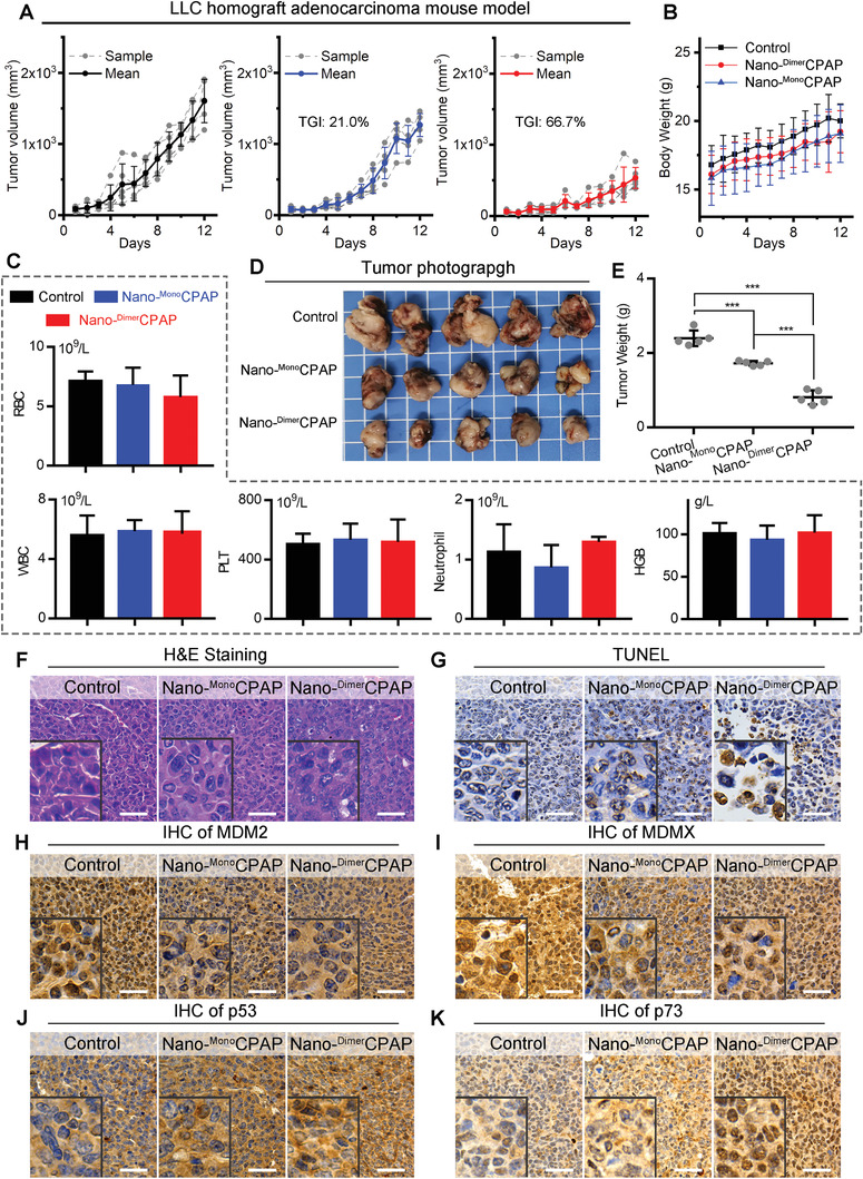
Figure 3.

Nano‐DimerCPAP potently suppressed tumor growth superior to Nano‐MonoCPAP in C57/B6 mice LUAD allograft model. A) Growth curves of LLC homograft model in C57/B6 mice with treatments, following the administration of control (PBS), Nano‐DimerCPAP (2.5 mg kg−1) and Nano‐MonoCPAP (2.5 mg kg−1) (n = 5). B) Body weight of mice with the indicated treatments. C) Analysis of red blood cell (RBC), white blood cell (WBC), platelets (PLT), neutrophil, hemoglobin (HGB) in mice whole blood after treatments. D) Representative photographs and E) weight of tumor tissue isolated at the end of experiment. F,G) Representative images of H&E and TUNEL staining in tumor section from mice (scale bar: 50 µm). The immunohistochemical (IHC) staining for H) MDM2, I) MDMX, J) p53, and K) p73 in tumor sections from mice (scale bar: 50 µm).
