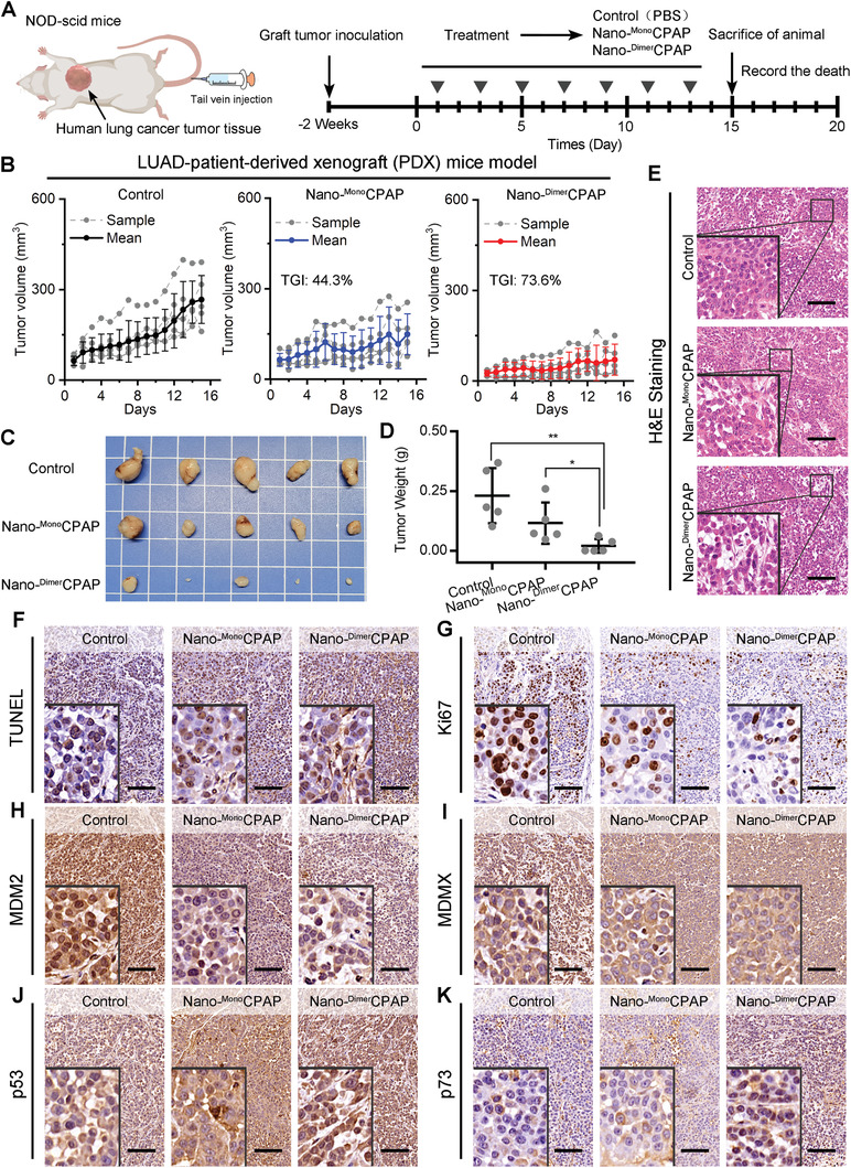
Figure 4.

Nano‐DimerCPAP enhanced more anticancer activity in LUAD patient‐derived tumor xenograft in NOD/SCID mice. A) Diagrammatic sketch of LUAD‐PDX mice model with indicated treatments. B) Growth curves of LUAD‐PDX mice model after administration of control (PBS), Nano‐DimerCPAP (2.5 mg kg−1) and Nano‐MonoCPAP (2.5 mg kg−1) (mean ± sd, n = 5 per group). C) Images and D) weights of tumors excised at the end of treatment. p‐values were calculated by t test (*p < 0.05; **p < 0.01; ***p < 0.001). E) The H&E and F) TUNEL staining in tumor from mice after indicated treatments (scale bar: 200 µm). Representative images of IHC staining of G) Ki67, H) MDM2, I) MDMX, J) p53, and K) p73 in tumor section from mice with indicated treatments (scale bar: 200 µm).
