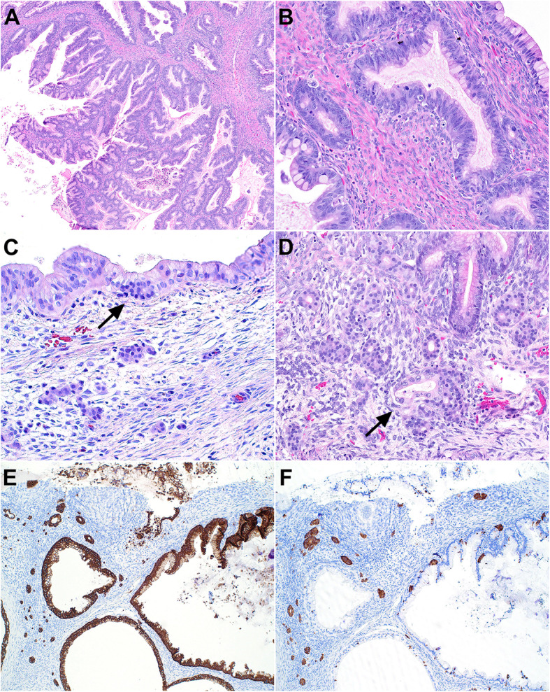
Fig. 1.

A, The cysts are lined by gastrointestinal-type mucinous epithelium with variable degrees of epithelial proliferation. B, Higher magnification demonstrating mucinous epithelium with goblet cells and nuclear atypia. C, There are several foci of bland, monotonous epithelioid cells arranged in solid nests or tubular/acinar architecture in reactive stroma adjacent to mucinous epithelium. The bland epithelioid cells can also be seen in adjacent mucinous glands (arrow). D, Florid stromal micronests are seen focally and cells emanating from a mucinous gland are present (arrow). E, Pancytokeratin (AE1/AE3) immunostaining highlights both mucinous epithelium and the stromal micronests. F, Synaptophysin immunostaining highlights the endocrine cell micronests in the stroma as well as intraepithelial neuroendocrine cells in adjacent mucinous glands
