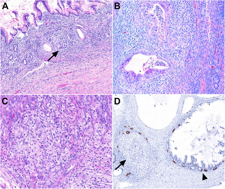
Fig. 2.

A, Endocrine cell micronests are seen in stroma adjacent to the mucinous cysts and their outpouching glands (arrow). B, Mucinous glands in early stage of degeneration with intraluminal neutrophils and stroma with reactive changes, histiocytes, and endocrine cell micronests. C, Round to ovoid shaped histiocytic infiltrates with endocrine cell micronests at the periphery, representing the late-stage glandular degeneration with preserved endocrine cells. D. Synaptophysin immunostaining highlights stromal endocrine cell micronests and intraepithelial neuroendocrine cells. A completely degenerated mucinous cyst with preserved neuroendocrine cells (arrow) is seen adjacent to a relatively intact mucinous cyst demonstrating intraepithelial neuroendocrine cells with variable degrees of proliferation (arrowhead)
