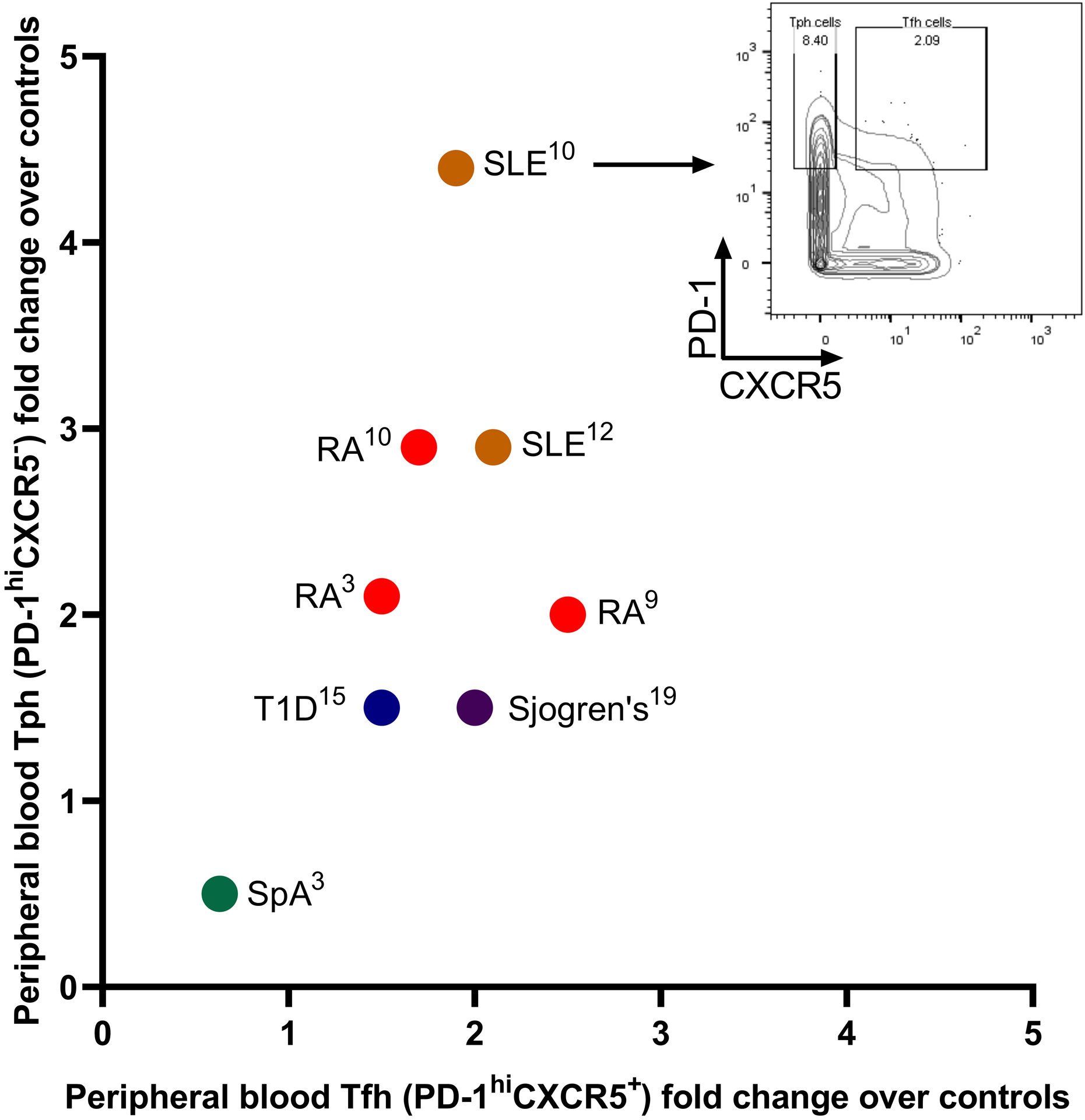
Figure 1.

Expansion of Tph cells and Tfh cells in autoimmune diseases. Tph cells and Tfh cells were considered as PD-1hiCXCR5− and PD-1hiCXCR5+, respectively, and are represented in each indicated disease as fold over controls according to the cited publications. Publications were included if they analyzed both Tph and Tfh cells in both the disease patients and controls and gated them by cytometry as PD-1hiCXCR5− and PD-1hiCXCR5+. Representative mass cytometry plot demonstrates peripheral blood memory CD4+ T cells from Bocharnikov et al.
