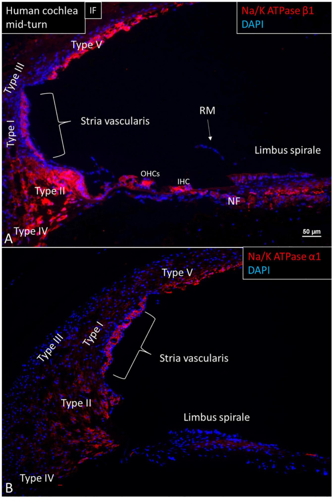
FIGURE 1.
Immunofluorescence (IF, A) and confocal microscopy (B) of cross-sectioned human cochlea shows expression of NKAβ1 and NKAα1. The marginal cells, type II, IV, and V fibrocytes, outer sulcus, Hensen cells, interdental cells, spiral limbus fibrocytes, and auditory neurons express NKAβ1 but not type I and III fibrocytes, RM and the tympanic covering layer. The NKAα1 subunit is strongly expressed in the SV marginal cells but less in type IV and V fibrocytes. OHCs, outer hair cells; IHC, inner hair cell.

