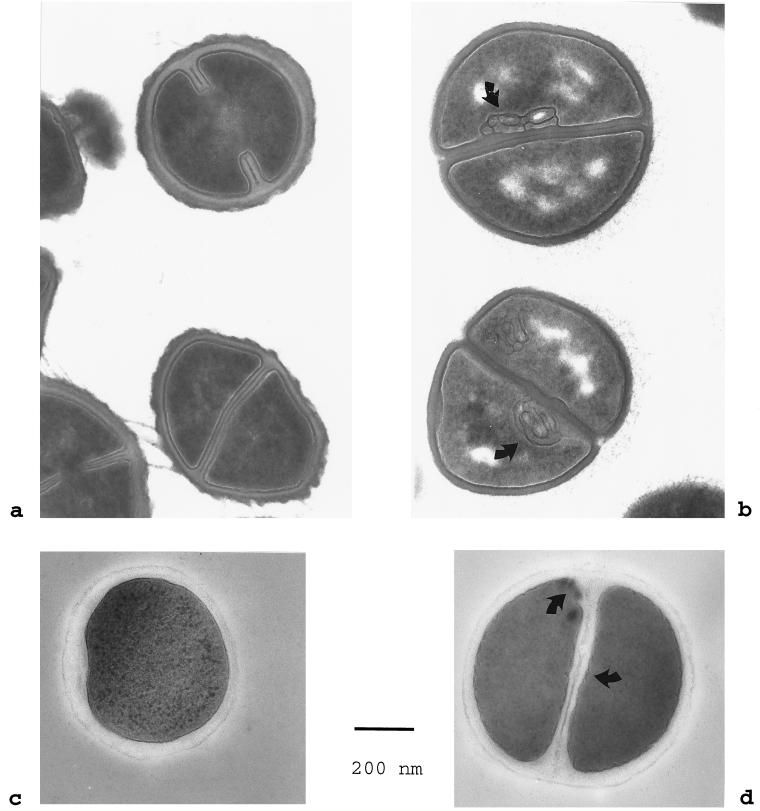
FIG. 4.
Transmission electron microscopy of S. aureus Smith diffuse. (a and c) Morphology of control bacteria incubated in phosphate-buffered saline for 120 min as seen after chemical fixation and after cryofixation and freeze-substitution, respectively; (b) prominent mesosome-like membrane infoldings (arrows) and segregation of the cytoplasm in chemically fixed specimens after 120 min of incubation in 50 μM NCT solution (pH 7.0); (d) undulations and slight infoldings of the bacterial cell membrane (arrows) after 120 min of incubation in NCT as seen in cryofixed, freeze-substituted samples. Bar = 200 nm. Magnification, ×62,500.

