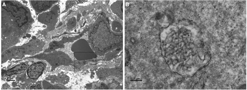
Figure 2.
Electron microscopy showing tubuloreticular inclusions in capillary endothelial cells that have been seen in COVID-19–related collapsing glomerulopathy.2 A, ×4200; (B) ×60 000. Electron microscopy images were obtained by Jesus Macias, senior histotechnologist and electron microscopist at University of California San Diego Health.

