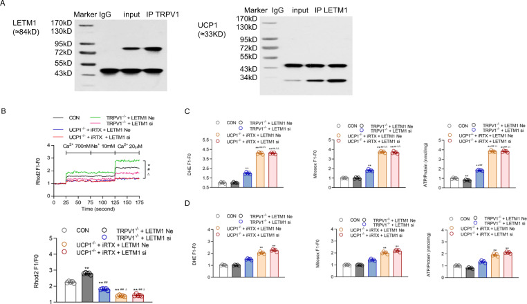
Fig. 6.
Mechanism by which TRPV1 and UCP1 double knockout cause mitochondrial ion imbalance. A Immunoprecipitation (IP) with TRPV1/LETM1 and LETM1/UCP1 antibodies on mitochondria isolated from the brown adipose tissue of WT mice. The immunoblot bands are representative of six separate experiments. B Mitochondrial [Ca2+] of primary cultures of brown adipocytes from WT, TRPV1−/− and UCP1−/− mice, as well as TRPV1−/− and UCP1−/− mice transfected with LETM1 siRNA, was measured after loading with Rhod2 (2 μmol/L). The lower panel is the bar graph derived from the left figure with [Ca2+] at 20 μM. C, D Dihydroetorphine hydrochloride (DHE, C/D, left panel) and ATP (C/D, right panel) levels of primary cultured brown adipocytes were measured before (C) and after tempol treatment (200 µM, D) treatment. MitoSox levels of primary cultured brown adipocytes were measured before (C, middle panel) and after mito-tempo (20 µM, D, middle panel) treatment. *P < 0.05, **P < 0.01 vs. WT mice (CON); ##P < 0.01 vs. TRPV1−/− mice + LETM1 siRNA negative control (TRPV1−/− + LETM1 Ne); ∆∆P < 0.01 vs. UCP1−/− mice + iRTX + LETM1 siRNA negative control (UCP1−/− + LETM1 Ne)

