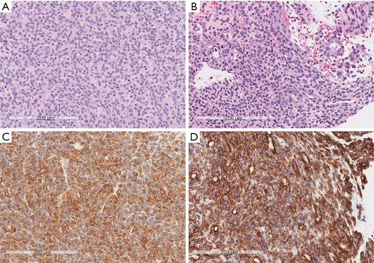
Figure 2.
Hematoxylin and eosin staining and IHC of tumor specimens. (A) Areas of round cells with a high-mitotic index. (B) Areas of round and spindled cells with a lower mitotic rate. (C) Positive immunohistochemical staining for SMA. (D) Positive immunohistochemical staining for collagen IV. Scale bar: 200 µm. IHC, immunohistochemistry; SMA, smooth muscle antigen.

