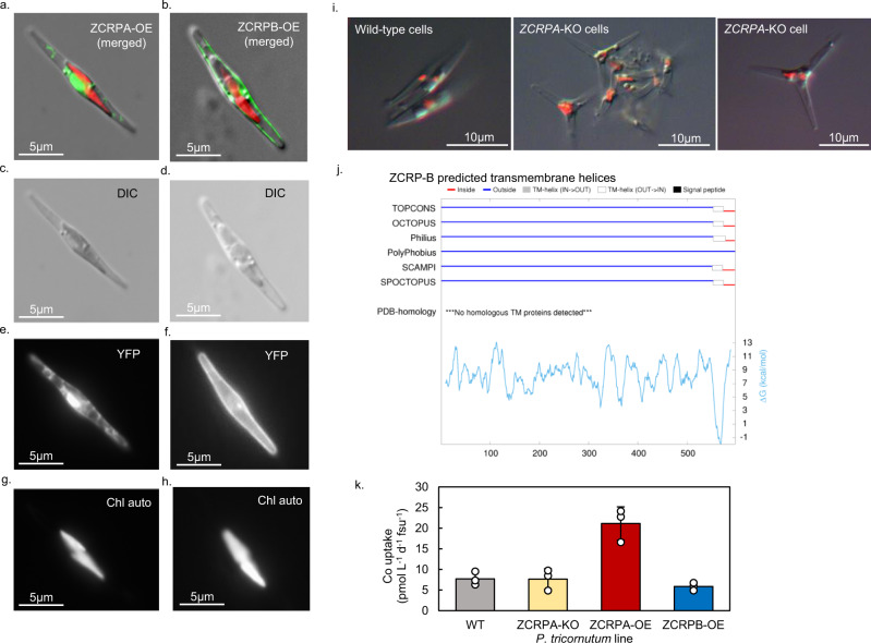
Fig. 4. Confocal and epifluorescent micrographs of the ZCRP-A and ZCRP-B proteins fused to YFP and overexpressed in P. tricornutum.
a Localization of ZCRP-A to the chloroplasts and b localization of ZCRP-B to the cell membrane. YFP fluorescence is false-colored green and chlorophyll autofluorescence is false-colored red. Composite images are stacks of the individual channels c, d differential interference contrast (DIC), e, f yellow fluorescent protein (YFP), and g, h chlorophyll autofluorescence (Chl auto). i Micrographs of Zn-limited wild-type and ZCRP-A knockout (KO) P. tricornutum cells showing morphological differences. For a–h and i, results were validated > 10 times. j Topology predictions from five sub-methods (OCTOPUS, Philius, PolyPhobius, SCAMPI, and SPOCTOPUS), consensus prediction (TOPCONS), and predicted ΔG values for P. tricornutum ZCRP-B generated using the TOPCONS webserver (https://topcons.cbr.su.se/)27,28. k Extent of Co uptake after 24 h for wild-type (WT), ZCRPA-knockout (KO), and ZCRPA-overexpression (OE) lines of P. tricornutum normalized to fluorescence units (fsu). Data are presented as mean values ± the standard deviation of biological triplicate cultures (n = 3). Individual data points are overlaid as white circles. The extent of Co uptake was found to be significantly larger in the ZCRPA-OE line compared to the wild-type via one-way ANOVA (f(3) = 23.16, p = 0.000268) and post hoc Dunnett test (p = 0.00048).

