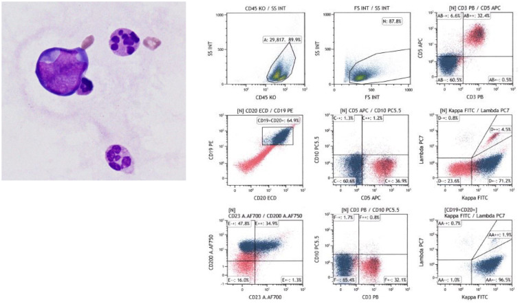
Fig. 2.
Cytological examination of the pericardial fluid and flow cytometry analysis
May-Giemsa staining shows abnormal cells with an irregularly shaped nucleus (left, ×1000). Flow cytometry analysis revealed that the lymphoid cells were positive for CD19, CD20, and immunoglobulin kappa, and negative for CD3, CD5, and CD10.

