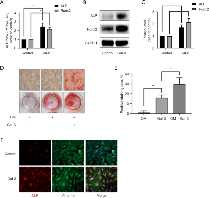
Figure 2.
Effect of Gal-3 on calcification-related gene/protein expression and osteogenic differentiation of hVICs. (A) hVICs interfered with or without 2 µg/mL Gal-3 for 72 h, and the mRNA expression levels of ALP and Runx2 detected by qRT-PCR (n=4); (B) Western blotting of Gal-3-treated ALP and Runx2 proteins; (C) quantification of western blot (n=4); (D) Alizarin Red S staining of the cells under different conditioned treatments, including control (normal culture medium), OM (osteogenic medium), and OM + Gal-3 (osteogenic medium with Gal-3 treatment). The microscopic images were taken with the 4× objective; (E) quantification of alizarin red positive area (n=3); (F) immunofluorescent staining was carried out to detect ALP in hVICs treated with or without Gal-3 (scale bar: 50 µm). Statistical comparisons were made using Student’s t-test. All of the data are presented as mean ± SEM. *, P<0.05, **, P<0.01. Gal-3, galectin-3; hVICs, human aortic valve interstitial cells; qRT-PCR, quantitative real-time polymerase chain reaction.

