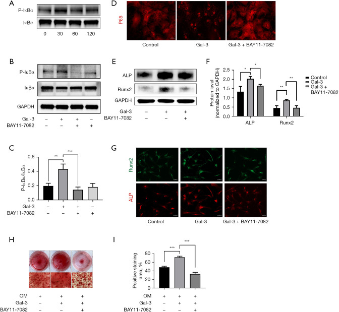
Figure 4.
Gal-3 treatment activated NF-κB signaling pathway in hVICs. (A) Western blot analysis revealed the phosphorylation level of IκBα in hVICs treated with Gal-3 for 0, 30, 60, and 120 min; (B) the phosphorylation level of IκBα in hVICs treated with or without Gal-3 or BAY11-7082 for 30 min, assessed by Western blot; (C) quantification of western blot; (D) immunofluorescence of p65 in hVICs treated with or without Gal-3 or BAY11-7082 for 30 min; (E) the protein expression levels of osteogenesis-specific markers (ALP, Runx2) were determined by Western blot in hVICs treated with OM, OM + Gal-3, or OM, Gal-3 + BAY11-7082 for 48 h; (F) quantification of western blot (n=3); (G) the immunofluorescence staining of ALP and Runx2 in AVICs with Gal-3 or Gal-3 + BAY11-7082; (H) representative images of Alizarin Red staining showing calcium deposits in hVICs treated with OM, OM + Gal-3 or OM, Gal-3 + BAY11-7082 for 21 days. The microscopic images were taken with the 4× objective; (I) quantification of alizarin red positive area (n=3). Statistical comparisons were made using Student’s t-test. All of the data are presented as mean ± SEM. *, P<0.05, **, P<0.01, ***, P<0.001. Gal-3, galectin-3; hVICs, human aortic valve interstitial cells.

