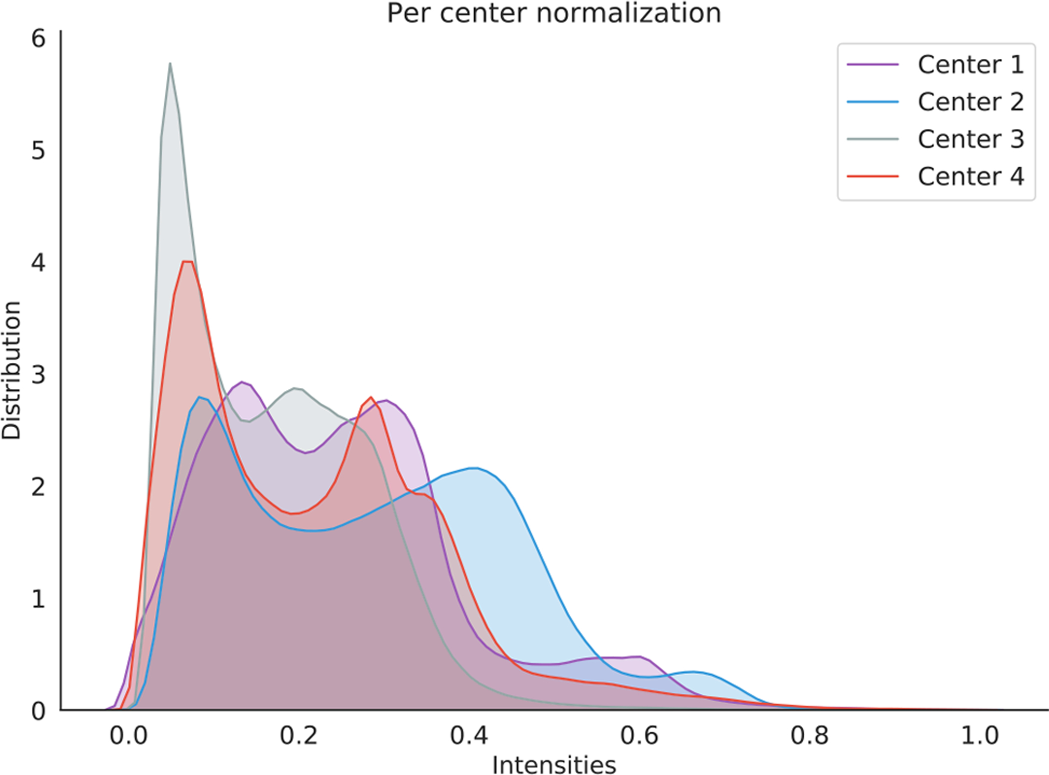
Fig. 2.

Intensity distribution of MRI axial-slice pixels from four different datasets (i.e., UCL, Montreal, Zurich, and Vanderbilt) that collected for gray matter segmentation. Intensity is normalized between 0 and 1 for each site. Image courtesy to C. Perone [25].
