Figure 3.

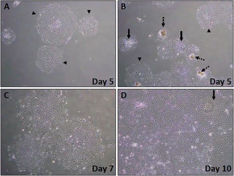
Phase‐contrast images showing early stages of iPSC‐K differentiation (days 5‐10). (A) Healthy colonies at day 5 after start of iPSC‐K differentiation (arrowheads). (B) Mix of healthy and unhealthy colonies at day 5 of differentiation. Dashed arrows point to colonies that did not properly attach to the plate. Note the darker (brown) cell clumps. These colonies will die. Solid arrows point to colonies that do not look ideal, but that may differentiate into keratinocytes. Arrowheads point to colonies with the expected epithelial morphology. (C) Expanding early‐stage differentiating colony at day 7. (D) Higher magnification of a day 10 colony. Note the emerging cobblestone appearance, a typical feature of epithelial cells. Arrow points to cells that have acquired the expected cobblestone morphology.
