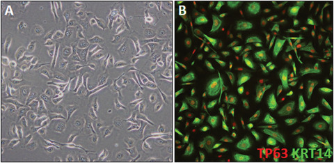
Figure 5.

Fully differentiated iPSC‐K. (A) Phase‐contrast image of day 45 iPSC‐K. The cells show typical cobblestone keratinocyte morphology. A few cells appear to differ in morphology. These cells are mostly likely migratory leading to the elongated appearance. (B) Example of a day 43 culture labelled with TP63 and KRT14 antibodies.
