Figure 6.

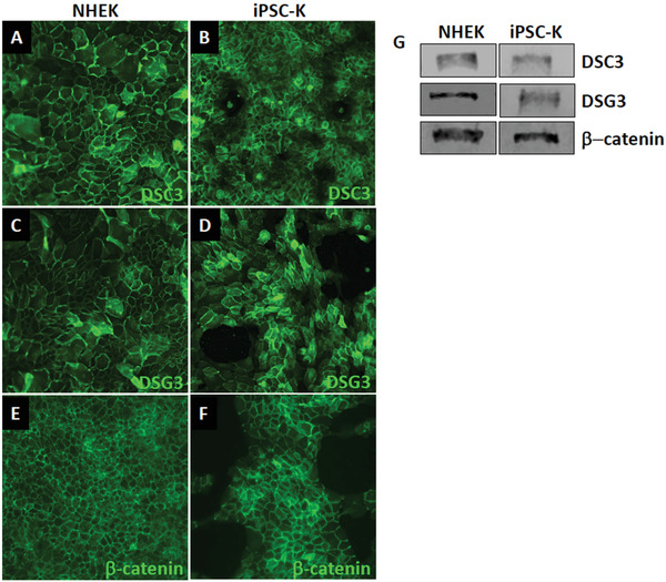
Expression of keratinocyte markers in iPSC‐K. iPSC‐K and primary human epidermal keratinocytes were exposed to 1.3 mM Ca2+ for 48 hr. (A‐F) Immunofluorescence staining and (G) Western blot analysis demonstrates normal expression and localization of desmosomal proteins (DSC3, DSG3) and an adherens junction protein (β‐catenin). Antibodies used for immunofluorescence staining are: DSG3 (clone 5H10; courtesy of Dr. James K Wahl III, PhD, University of Nebraska Medical Center, Lincoln, NE), DSC3 (Progen cat. no. 61093), and β‐catenin (Santa Cruz cat. no. sc‐7963). Antibodies used forlotting are: DSG3 (Invitrogen cat. no. MA5‐16025), DSC3 (Progen cat. no. 61093), and β‐ catenin (Santa Cruz cat. no. sc‐7963).
