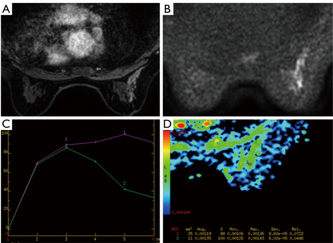
Figure 1.
MRI imaging in a 45-year-old woman. (A) Contrast enhanced breast MRI showed segmental distribution and clustered-ring enhancement; (B,C) the lesion showed diffusion restriction on DWI and wash-out dynamic curve; (D) the ADC value of this malignant lesion equals to 1.19×10⁻³ mm2/s. Final biopsy showed invasive ductal carcinoma. MRI, magnetic resonance imaging; DWI, diffusion-weighted images; ADC, apparent diffusion coefficient.

