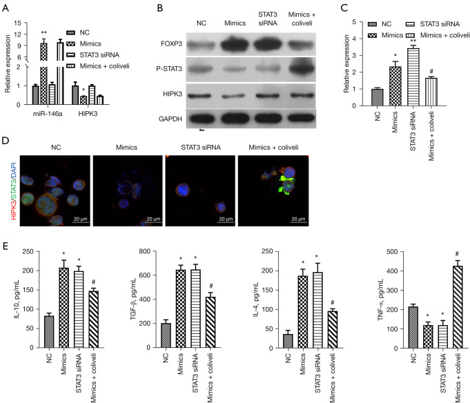
Figure 5.
Activation of STAT3 prominently reversed miR-146a-mediated inhibition of p-STAT3 and inflammation in TGF-β-induced thymocytes. TGF-β-induced thymocytes were transfected or treated with NC, miR-146a mimics, STAT3 siRNAs or miR-146a mimics + colivelin. (A) The relative expressions of miR-146a and HIPK3 were examined by RT-qPCR in the treated thymocytes. (B) Western blot analysis of expression of HIPK3, FOXP3 and p-STAT3 in each group. (C) Expression level of FOXP3 was assessed by RT-qPCR. (D) The co-localization and expression of HIPK3 and STAT3 were examined through IF assay. Magnification, 400×, scale bar =20 µm. (E) ELISA assay was carried out to evaluate the levels of IL-10, TGF-β, IL-4, and TNF-α in each group of thymocytes. *P<0.05, **P<0.01 vs. NC group; #P<0.05 vs. mimics group. NC, negative control; siRNA, small interfering RNA; HIPK3, homeodomain-interacting protein kinases 3; IL-10, interleukin 10; TGF-β, transforming growth factor-β; IF, immunofluorescence; IL-4, interleukin 4; TNF-α, tumor necrosis factor-α; RT-qPCR, reverse transcription quantitative polymerase chain reaction; ELISA, enzyme-linked immunosorbent assay.

