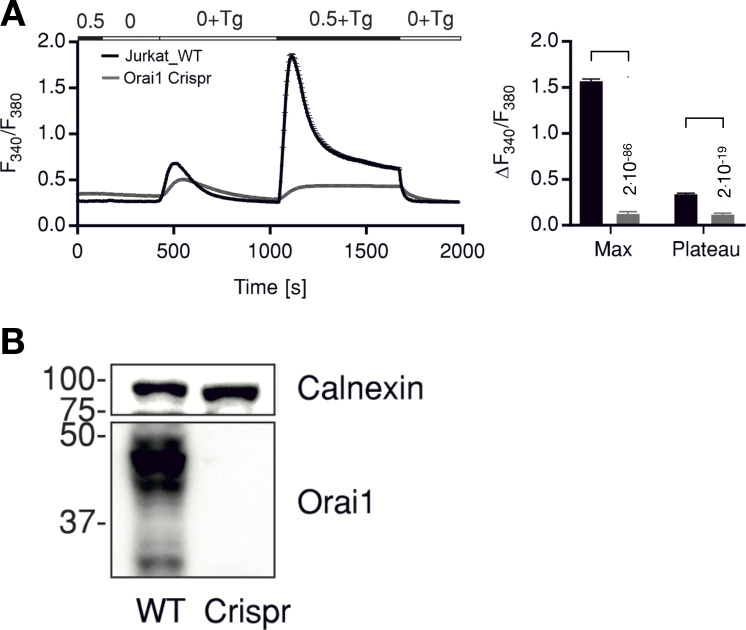
Figure S1.
Knockout of Orai1 in Jurkat cells abolishes Tg induced Ca2+ release. (A) Average traces showing changes in Ca2+cyt as indicated by fluorescence ratio (F340/F380) of ratiometric calcium dye Fura-2 over time in response to Tg-induced ER calcium store depletion and Ca2+ readdition by changing extracellular solutions as shown in the bar above. Measurements were done in control Jurkat cells (WT, black) or in cells where Orai1 was deleted (Orai1-Crispr, gray). The right panel shows quantification of maximum (Max) and steady-state (Plateau) changes in fluorescence ratio + SD from 135 to 176 cells measured in N = 3 independent experiments. Statistical differences between WT and Crispr cells were analyzed by unpaired Student’s t test, and respective P values are given in the figure. (B) Western blot analysis of cells measured in A showing deletion of Orai1 in Crispr-Cas9–treated cells. Source data are available for this figure: SourceData FS1.

