
FIGURE 1.
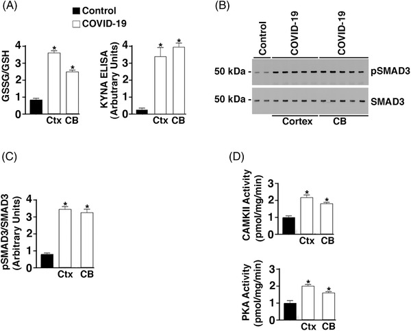
Increased oxidative stress, inflammatory and adrenergic signaling in brains of COVID‐19 patients. A, Bar graph depicting the glutathione disulfide (GSSG)/ glutathione (GSH) ratio and kynurenic acid (KYNA) enzyme‐linked immunsorbent assay signal from control (n = 6) and COVID‐19 (n = 6) tissue lysates. CB, cerebellum; Ctx, cortex. Data are mean ± standard deviation (SD). *P < .05 control versus COVID‐19. B, Western blots showing phospho‐SMAD3 and total SMAD3 from control (n = 4) and COVID‐19 (n = 7) brain lysates. C, Bar graphs depicting quantification of pSMAD3/SMAD3 from Western blot signals in B. D, Calmodulin‐dependent protein kinase II association domain (CaMKII) and protein kinase A (PKA) activity of brain tissue lysates. Data are mean ± SD. *P < .05 control versus COVID‐19