
FIGURE 2.
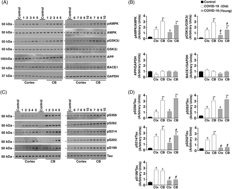
Hyperphosphorylation of tau but normal amyloid precursor protein (APP) processing in COVID‐19 brains. A, Brain (CB, cerebellum; Ctx, cortex) lysates were separated by 4% to 20% polyacrylamide gel electrophoresis. Immunoblots were developed for pAMPK, AMPK, GSK3β, pGSK3β (T216), APP, BACE1, and GAPDH loading control. The numbers (1–10) above immunoblots refer to patient numbers listed in Table 1. B, Bar graphs showing quantification of pAMPK, pGSK3β, APP/GAPDH, and BACE1/GAPDH from Western blots in (A). Data are mean ± standard deviation (SD). *P < .05 control versus COVID‐19; **P < .05 CB versus Ctx; #P < .05 COVID (Young) versus COVID (Old). C, Immunoblots of brain lysates showing total tau and tau phosphorylation on residues S199, S202/T205, S214, S262, and S356. D, Bar graphs showing quantification phosphorylated tau at the residues shown on Western blots in (C). Data are mean ± SD. *P < .05 control versus COVID‐19; **P < .05 CB versus Ctx; #P < .05 COVID (Young) versus COVID (Old)