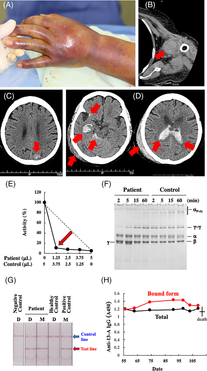
FIGURE 1.

The major bleeding symptoms/sites on day 55 (A–C) and day 109 (D); the results of an experimental work‐up for AiF13D on day 56 (E–H). (A) Ecchymosis involving the left hand and forearm. She had pain and numbness in her fingers. (B) Computed tomography scan showing her left shoulder joint bleeding. (C) central nervous system (CNS) bleeding in her left posterior parietal lobe. (D) Computed tomography scan showing right subcortical and subarachnoid CNS hemorrhages, that had perforated into the ventricles. She also had a subcutaneous hematoma on the right side of the head. (E) The five‐step dilution cross‐mixing test by an amine incorporation assay was performed using the patient's plasma at the ratios of 0:1, 1:3, 1:1, 3:1, and 1:0 with a normal plasma. The mixed samples were incubated at 37°C for 2 h, before the assay. The patient's sample showed a downward concave “inhibitor” pattern. A straight broken line depicts a theoretical “deficient” pattern. (F) Fibrin cross‐linking reaction. Gamma‐chain dimerization was significantly retarded and γ‐chain monomer remained even after 5 min. Alpha‐chain polymerization was almost absent. (G) Immunochromatographic test for anti‐FXIII‐A subunit autoantibodies with (spiked; M) or without (direct; D) mixing patient's and normal control's plasma showed positive results. (H) Enzyme‐liked immunosorbent assay for anti‐FXIII‐A subunit autoantibodies (day 56–day 91). Despite the administration of Prednisolone and IVIg, the amounts of both F13‐bound (Bound Form) and total anti‐F13 autoantibodies in our case remained almost unchanged. Filled‐black and ‐gray circles depict bound forms and total (free and bound forms) amounts of anti‐FXIII‐A subunit IgG, respectively [Color figure can be viewed at wileyonlinelibrary.com]
