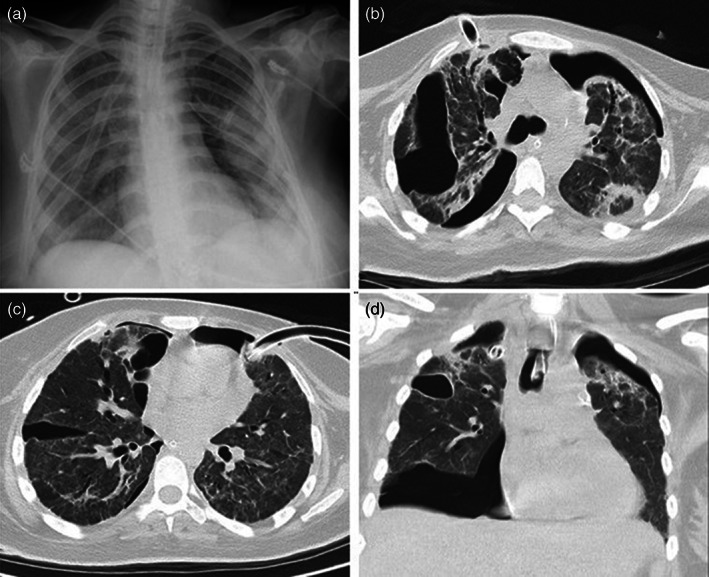
FIGURE 2.

Chest CT and X‐ray imaging of the patient. (a) In the posterior–anterior chest X‐ray imaging, drainage catheters in the upper lobes of both lungs and pneumothorax in the left lung are observed. (b) In the axial chest CT image parenchymal window, pneumothorax in both lungs and consolidated areas in the lung parenchyma, interlobular thickening, bronchiectasis changes, and a drain catheter in the anterior upper lobe of the right lung are observed. (c) In the axial chest CT image parenchymal window, pneumothorax in both lungs, and a drain catheter in the left lung upper lobe anterior are observed. (d) In the axial chest CT image parenchymal window, an intubation tube is observed in the trachea with pneumothorax in both lungs and consolidations in both upper lobes
