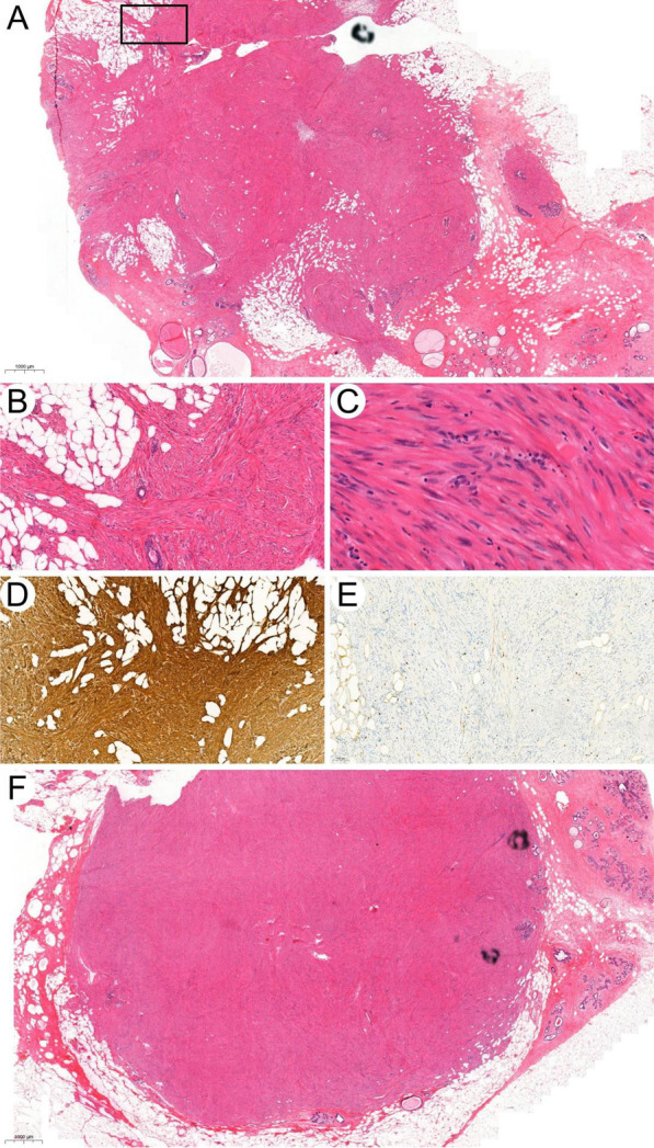
Fig. 2.

Bilateral leiomyoma of breast. A Leiomyoma of left breast. Noted the invasive growth pattern of this tumor including its infiltrative margin and the packed normal mammary glands in it. The infiltrative margin indicated by the black square is magnified in B (H&E, × 10). B Noted the infiltration of this tumor into adipose tissue (H&E, × 200). C Nuclei of tumor cells in high magnificent view showed typical spindle morphology with blunt end which is characteristics for leiomyoma (H&E, × 400). D, E Immunohistochemical stain of SMA and Ki-67 in leiomyoma of left breast. F Leiomyoma of right breast (H&E, × 10)
