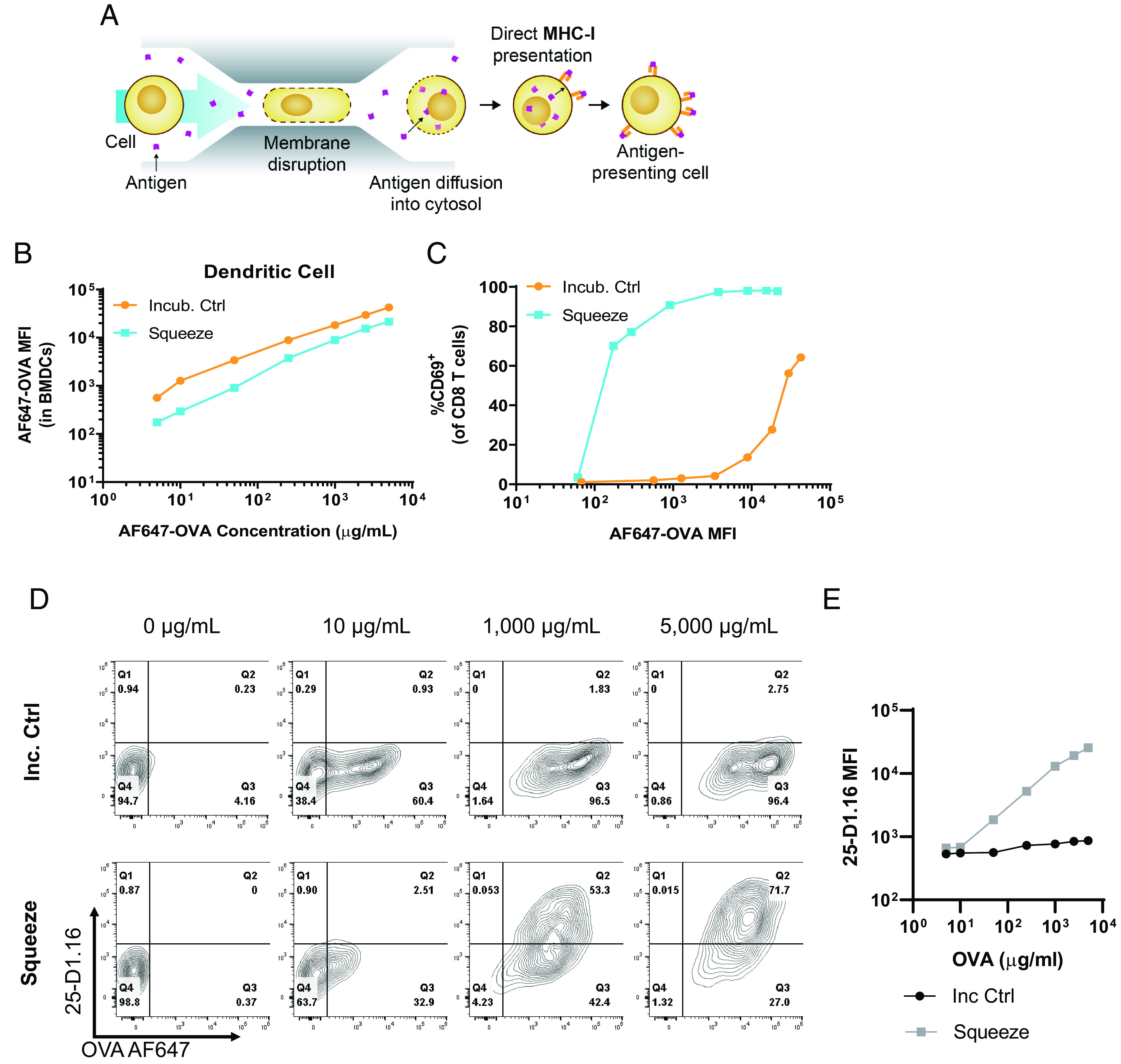
FIGURE 1.

Microfluidic squeezing enhances Ag presentation by professional APCs. (A) Cells are squeezed in the presence of Ag in a microfluidic chip that creates temporary pores in the cells, allowing Ag to diffuse into the cytosol, from which it can be presented on MHC-I. (B–E) Mouse BMDCs were incubated with varying concentrations of fluorescently labeled OVA for 30 min at 37°C (Incub. Ctrl) or squeezed with the same concentrations (Squeeze). (B) OVA MFI for Incub. Ctrl and Squeeze groups at varying OVA concentrations. (C) Incub. Ctrl and Squeeze BMDCs were cocultured with OT-I CD8+ T cells for 24 h, and CD69 staining on the OT-I cells was assessed by flow cytometry. (D and E) Mouse BMDCs were incubated with varying concentrations of fluorescently labeled OVA for 30 min at 37°C (Incub. ctrl) or were squeezed with the same concentrations (Squeeze). Incub. ctrl and squeezed BMDCs were incubated for 4 h at 37°C and subsequently stained with 25-D1.16 Ab and analyzed by flow cytometry. Representative plots for indicated concentrations are shown in (D). Summary data for all concentrations are shown in (B). Data are representative of two independent experiments.
