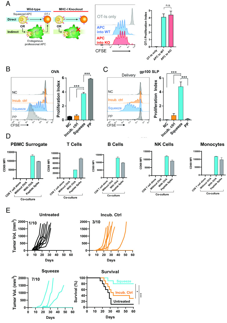
FIGURE 3.
Ag presentation by squeezed immune cells is direct and primes antitumor immunity. (A) A total of 2.5 × 10⁶ CFSE-labeled CD8+ OT-I cells were transferred into either WT or MHC-I−/− (knockout [KO]) mice. A few hours later, 5 × 10⁶ splenocytes squeezed in the presence of OVA and incubated for 4 h with CpG were injected i.v. After 3 d, lymph nodes were harvested, and proliferation of OT-I cells was assessed by CFSE dilution. n = 5 mice per group. (B) Mouse B cells were not in contact with OVA (NC), incubated at room temperature with OVA (Incub. ctrl), squeezed in the presence of OVA (Squeeze), or pulsed with SIINFEKL peptide for 1 h at 37°C. A total of 5 × 10⁶ B cells were coinjected with 3 µg LPS to immunize mice that had also received 2.5 × 10⁶ CFSE-labeled OT-I CD8+ T cells. CFSE dilution by the OT-I cells was assessed 3 d after immunization in lymph nodes. n = 5 mice per group. (C) Mouse B cells were not in contact with gp100 SLP (NC), incubated at room temperature with gp100 SLP (Incub. ctrl), squeezed in the presence of the gp100 SLP (Squeeze), or pulsed with short peptide for 1 h at 37°C (PP). A total of 5 × 10⁶ B cells were coinjected with 3 µg LPS to immunize mice that had also received 2.5 × 10⁶ CFSE-labeled pmel CD8+ T cells. CFSE dilution by the pmel CD8+ T cells was assessed 3 d after immunization. n = 5 mice per group. (D) Mouse splenocytes were squeezed with or without OVA. After squeezing, indicated populations of cells were magnetically separated and cocultured with OT-I TCR-transgenic cells overnight. CD69 MFI on OT-I cells was assessed by flow cytometry. Data are representative of two independent experiments. (E) Mice were left untreated or immunized on day −14 and day −7 with 1 × 10⁶ murine PBMC surrogate cells incubated or squeezed with OVA protein and matured with CpG. On day 0, mice were s.c. implanted with E.G7-OVA and monitored for tumor growth. ***p < 0.001.

