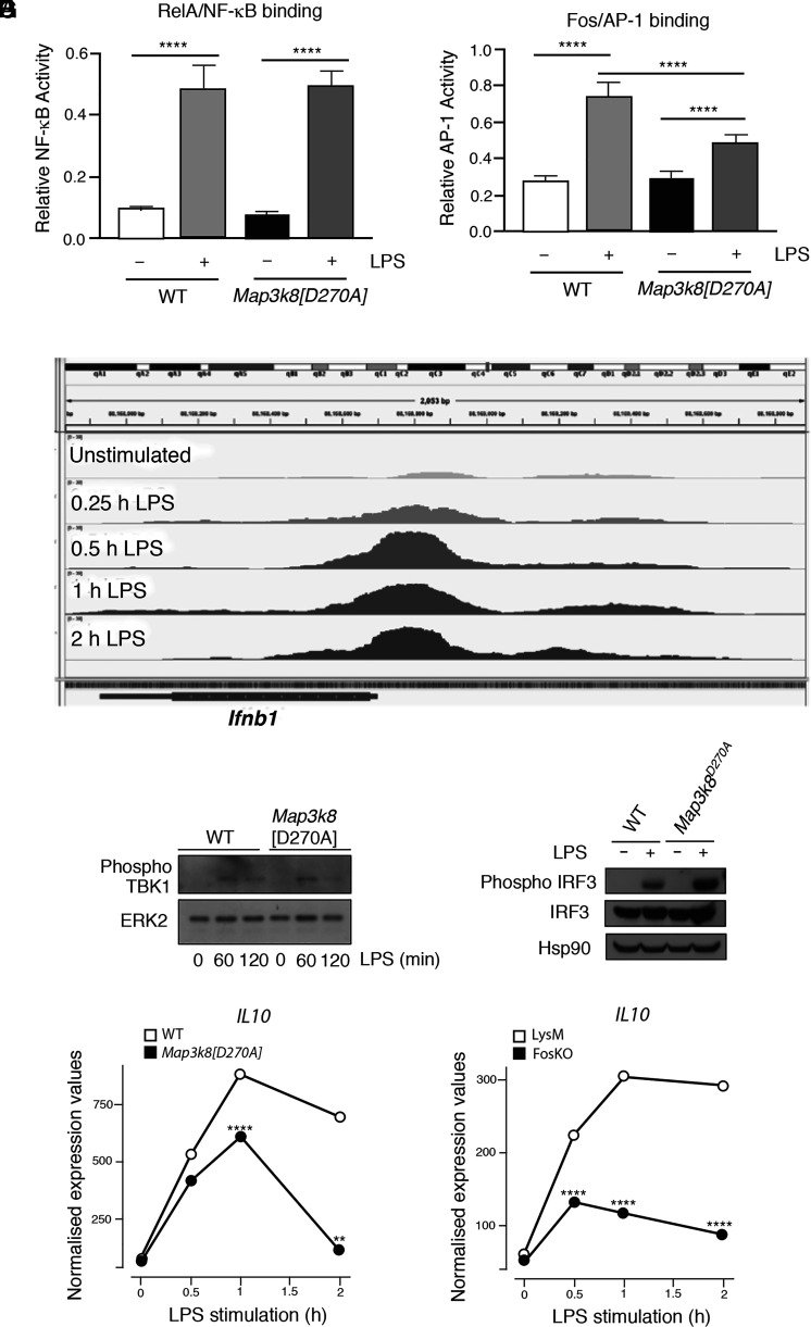
FIGURE 6.
TPL-2 regulation of Ifnb1 mRNA expression via multiple pathways. (A and B) WT and Map3k8[D270A] BMDMs were stimulated with LPS (30 min). Nuclear extracts were assayed by ELISA for RelA binding to NF-κB and FOS binding to AP-1. Mean of separate cultures from four biological replicates shown ± SEM. ****p < 0.0001. Two-way ANOVA. Representative of three experiments with similar results. (C) ChIP-seq data from Tong et al. (46) of FOS binding to Ifnb1 gene in BMDMs stimulated with LPS during a time course. The figure is an annotated screenshot of the Integrative Genomics Viewer output at the Ifnb1 gene location within the mm10 genome. (D and E) WT and Map3k8[D270A] BMDMs were stimulated with LPS at the indicated times in (D) and for 60 min in (E). Total lysates were immunoblotted for the indicated antigens. (F) Normalized counts from the RNA-seq analysis of WT and Map3k8[D270A] macrophages unstimulated or stimulated with LPS for the indicated times are shown Il10. **p ≤ 0.01, ****p ≤ 0.0001. (G) Normalized counts for Il10 from the RNA-seq analysis of FosKO and LysM-Cre BMDMs stimulated with LPS for the indicated times. ****p ≤ 0.0001.

