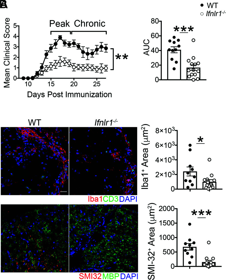
FIGURE 1.
Loss of IFNL signaling reduces severity of CNS autoimmune disease. (A) EAE clinical course and area under the curve (AUC) analysis following active immunization with MOG35-55 peptide of WT and Ifnlr1−/− mice. Data are pooled from two independent experiments; n = 11 WT and n = 14 Ifnlr1−/− animals shown. (B) IF analysis of lesions within the ventral lumbar spinal cord of WT and Ifnlr1−/− mice during EAE at chronic time point indicated in (A). Iba1+ and SMI-32+ areas were quantified. Data are pooled from two independent experiments; n = 10 WT and n = 14 Ifnlr1−/− animals shown. Scale bar, 20 μm. Data are presented as means ± SEM. *p < 0.05, **p < 0.01, ***p < 0.001 by Mann-Whitney U test (A) and two-tailed Student t test (A and B).

