FIGURE 4.
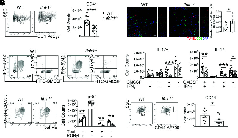
IFNL signaling maintains Th1 cell effector function during acute EAE. (A and B) Activated MOG-specific Th1 clones were injected into naive WT or Ifnlr1−/− mice. Flow cytometric analysis of infiltrating cells was performed on lumbar spinal cords during peak EAE (Fig. 2A). CD4+ cells were identified from a live, single cell gate and quantified. Data are pooled from two independent experiments; n = 10 WT and n = 12 Ifnlr1−/− animals shown. (C) IF analysis of TUNEL+ and CD3+ colocalization in lumbar spinal cords at chronic EAE time point (Fig. 2A). Data shown for n = 8 WT and n = 7 Ifnlr1−/− animals. Scale bar, 20 μm. (D and E) Of the CD4+ cells shown in (B), GM-CSF+, IFN-γ+, and IL-17+ cells were identified and quantified. Data are pooled from two independent experiments; n = 10 WT and n = 12 Ifnlr1−/− animals shown. (F) CD4+ cells were gated for Tbet and RORγt and quantified. Data are representative of two independent experiments; n = 4 WT and n = 3 Ifnlr1−/− animals shown. (G) CD4+ cells were gated on CD44 and quantified. Data are pooled from two independent experiments; n = 9 WT and n = 10 Ifnlr1−/− animals shown. Data are presented as means ± SEM. *p < 0.05, **p < 0.01, ***p < 0.001, ****p < 0.0001 by two-tailed Student t test, comparing WT versus Ifnlr1−/−.

