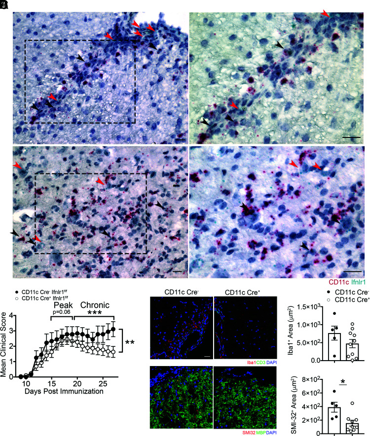
FIGURE 6.
CD11c+ cells are critical targets of IFNL during EAE. (A) Representative images of dual in situ hybridization for Ifnlr1 and CD11c RNA on WT spinal cords collected at peak EAE. Black arrowheads demonstrate colocalization of Ifnlr1 and CD11c. Red arrowheads depict Ifnlr1+CD11c− cells. Images on right are magnified images of the field of view shown on the left. (B) Ifnlr1fl/flCD11c-Cre− and Ifnlr1fl/flCD11c-Cre+ mice were monitored for EAE disease course following active immunization. Data are pooled from three independent experiments; n = 9 Ifnlr1fl/flCD11c-Cre− and n = 15 Ifnlr1fl/flCD11c-Cre+ animals shown. IF analysis of Iba1+ (C) and SMI-32+ area (D) in ventral lumbar spinal cord of Ifnlr1fl/flCD11c-Cre− and Ifnlr1fl/flCD11c-Cre+ mice at 27 d postimmunization. Scale bar, 20 μm. Data shown for n = 5 Ifnlr1fl/flCD11c-Cre− and n = 10 Ifnlr1fl/flCD11c-Cre+ animals (C) and n = 5 Ifnlr1fl/flCD11c-Cre− and n = 9 Ifnlr1fl/flCD11c-Cre+ animals (D). Data are presented as means ± SEM. *p < 0.05, **p < 0.01, ***p < 0.001 by Mann-Whitney U test (B) and two-tailed Student t test (C and D).

