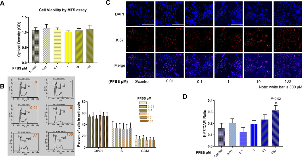
Fig 1. PFBS promotes proliferation in HTR-8/SVneo cells in a dose-dependent fashion.
(1A) Cell viability of HTR-8/SVneo cells after PFBS (0, 0.01, 0.1, 1, 10, 100 µM) exposure as measured by MTS assay with an absorbance of 490 nm.
(1B) Cell cycle analysis in HTR-8/SVneo cells treated with PFBS (0, 0.01, 0.1, 1, 10, 100 µM) as measured by flow cytometry.
(1C) Representative immunofluorescent images showing DAPI, Ki-67, and DAPI+Ki-67 staining for HTR-8/SVneo cells treated with PFBS (0, 0.01, 0.1, 1, 10, 100 µM).
(1D) Ratio of Ki-67(+)/DAPI(+) in HTR-8/SVneo cells after PFBS (0, 0.01, 0.1, 1, 10, 100 µM) exposure.

