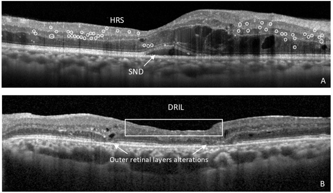
Fig. 1. OCT biomarkers of neuroinflammation and neurodegeneration in diabetic retinopathy (DR).
A Right eye of the patient with cystoid diabetic macular oedema (DMO) showing signs of neuroinflammation in the retina, such as subfoveal neuroretinal detachment (SND) and hyperreflective retinal spots (HRS), marked in circles. B Left eye of the patient with proliferative diabetic retinopathy (PDR) treated with numerous anti-VEGF intravitreal injections and vitrectomy, showing the presence of disorganization of the inner retinal layers (DRIL) and alterations in the integrity of outer retinal layers, in particular the external limiting membrane (ELM) and ellipsoid zone of the photoreceptors.

