FIGURE 3.

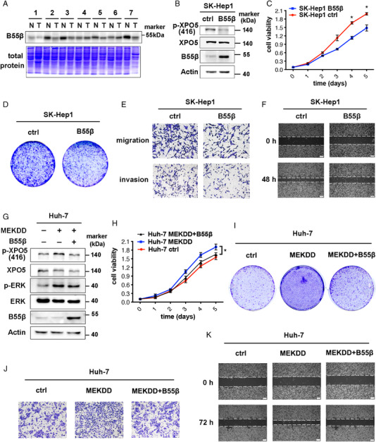
B55β inhibits growth, migration and invasion abilities of HCC cells. (A) The protein lysates of human HCC and adjacent normal tissues were subjected to SDS‐PAGE and then stained with coomassie blue or immunoblotted with anti‐B55β antibody. (B) B55β level was determined by Western blot in SK‐Hep1 stable cell lines with or without B55β overexpression. (C–F) MTT assays (C), cloning formation (D), transwell migration and invasion assays (E), and wound healing (F) for SK‐Hep1 B55β overexpression and ctrl cells. (G) B55β level was detected by Western blot in Huh‐7 stable cell lines with or without MEKDD/B55β overexpression. (H‐K) MTT assays (H), cloning formation (I), transwell migration assays (J), and wound healing (K) for Huh‐7 stable cell lines with or without MEKDD/B55β overexpression. Data are shown as the means ± SD. *p < 0.05.
