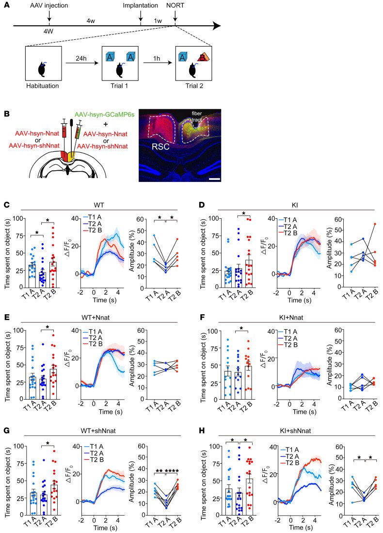
Figure 5. Abnormal neuronal activity is associated with the learning process in RSC of KI mice in short-term NORT.
(A) Schematic of experimental design. Viral injections were performed on 4-week-old mice, and fibers were implanted after a week. For the NORT, mice were exposed to 2 identical objects (object A) for 10 minutes on trial 1 (T1) after habituation. One hour later, one of the objects in A was replaced by a novel object (object B) for trial 2 (T2). (B) Left: schematic of viral injection into the RSC. Right: representative images of viral expression and location of optical fiber tract in RSC. Scale bar: 200 μm. (C–H) Left: exploring time for different objects. n = 18 for each group. Middle: average time courses of Ca2+ signal of different objects were shown for each group. Right: amplitudes of peak Ca2+ signal changes responding to different objects. (C and D) WT and KI control mice. n = 5 for calcium recording in each group. (E and F) Nnat-overexpressed WT and KI mice. n = 5 for calcium recording in each group. (G and H) Nnat-knockdown groups. n = 6 for calcium recording from WT mice injected with AAV-shNnat. n = 5 for calcium recording from KI mice injected with AAV-shNnat. Data are represented as mean ± SEM. *P < 0.05; **P < 0.01; ****P < 0.0001, paired t test.

