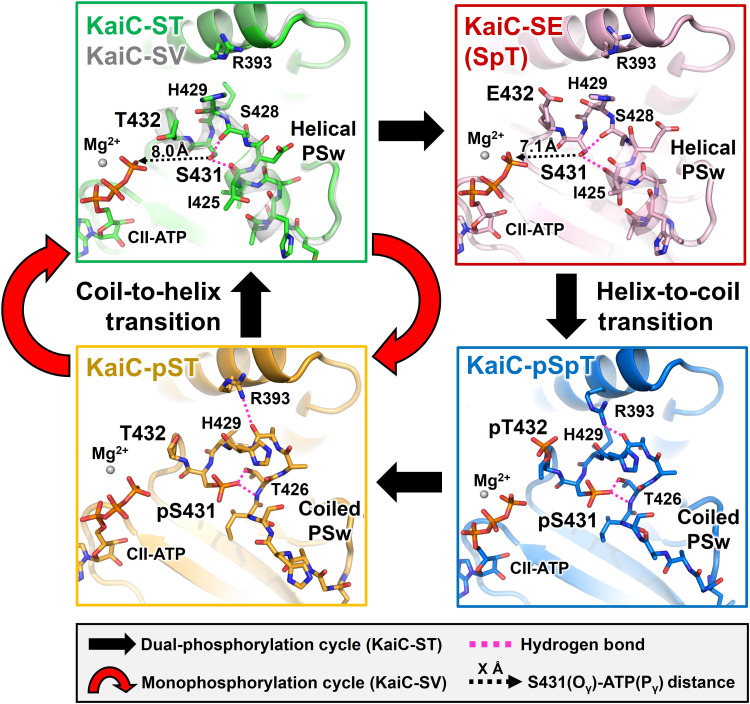
Fig. 2. Cyclic structural changes in the PSw located upstream of S431 in KaiC-ST (green), KaiC-SV (white), KaiC-SE (pink), KaiC-pSpT (blue), and KaiC-pST (orange).
Black and red arrows represent the dual-phosphorylation and monophosphorylation cycles in KaiC-ST and KaiC-SV, respectively. Dashed magenta lines correspond to hydrogen bonds around S431/pS431. Dashed black arrows indicate the distances between the S431 Oγ atom and CII-ATP Pγ atom.

