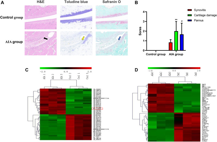
FIGURE 1.
Pathological and morphological changes and the different expression analysis of lncRNA and mRNA in synovial tissue from the control and AIA model group. (A) Compared with the control group, H&E staining of knee joint sections showed severe articular cartilage damage, adhesion and lymphocyte infiltration in the AIA group (black arrow); toluidine blue and safranin O staining showed that a large area of staining was lost and a large amount of chondrocytes was lost (yellow arrow), and bone damage was serious (blue arrow). (B) The quantitative analysis showed severe synovitis, cartilage damage and pannus of AIA model group (n = 3). Compared with controls, **p < 0.01, # p < 0.05. (C) Heat maps show the top 20 significantly upregulated and downregulated lncRNAs. lncRNA NR-133666 was enhanced in the AIA group. (D) Heat maps show the top 20 significant upregulated and downregulated mRNAs.

