. 2022 Feb 1;28(8):1507–1517. doi: 10.1158/1078-0432.CCR-21-3909

Figure 3.

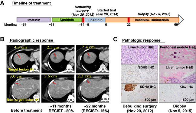
Figure 3. An example of durable treatment response in a patient with an SDH-deficient GIST. A, Treatment timeline and duration of various treatments the patient received for a metastatic SDH-deficient GIST. B, Representative CT images of the patient's metastatic liver lesions before, approximately 11 months and approximately 22 months after receiving the imatinib plus binimetinib combination therapy. One-dimension measurement in centimeters was provided for RECIST1.1 calculation at different time points. C, Treatment response by histopathologic studies. Representative images of histology and IHC stains for SDHB and SDHA, demonstrating dual SDH-deficiency, in the pretreatment tumor samples (debulking surgery; November 20, 2012) and the histology and the proliferation index marker, Ki67 IHC. The histology from the pretrial treatment liver lesions demonstrated more than 95% viable tumor and less than 5% treatment-associated necrosis. The histology from the on-treatment tumor samples (biopsy; November 5, 2015) showed 100% pathologic response with treatment-associated fibrosis, hyalinization, and dystrophic calcification in a resected metastatic peritoneal nodule and 70% pathologic response with treatment-associated necrosis in the metastatic liver lesion and less than 10% Ki67 IHC in the residual viable tumor. H&E, hematoxylin and eosin.

An example of durable treatment response in a patient with an SDH-deficient GIST. A, Treatment timeline and duration of various treatments the patient received for a metastatic SDH-deficient GIST. B, Representative CT images of the patient's metastatic liver lesions before, approximately 11 months and approximately 22 months after receiving the imatinib plus binimetinib combination therapy. One-dimension measurement in centimeters was provided for RECIST1.1 calculation at different time points. C, Treatment response by histopathologic studies. Representative images of histology and IHC stains for SDHB and SDHA, demonstrating dual SDH-deficiency, in the pretreatment tumor samples (debulking surgery; November 20, 2012) and the histology and the proliferation index marker, Ki67 IHC. The histology from the pretrial treatment liver lesions demonstrated more than 95% viable tumor and less than 5% treatment-associated necrosis. The histology from the on-treatment tumor samples (biopsy; November 5, 2015) showed 100% pathologic response with treatment-associated fibrosis, hyalinization, and dystrophic calcification in a resected metastatic peritoneal nodule and 70% pathologic response with treatment-associated necrosis in the metastatic liver lesion and less than 10% Ki67 IHC in the residual viable tumor. H&E, hematoxylin and eosin.