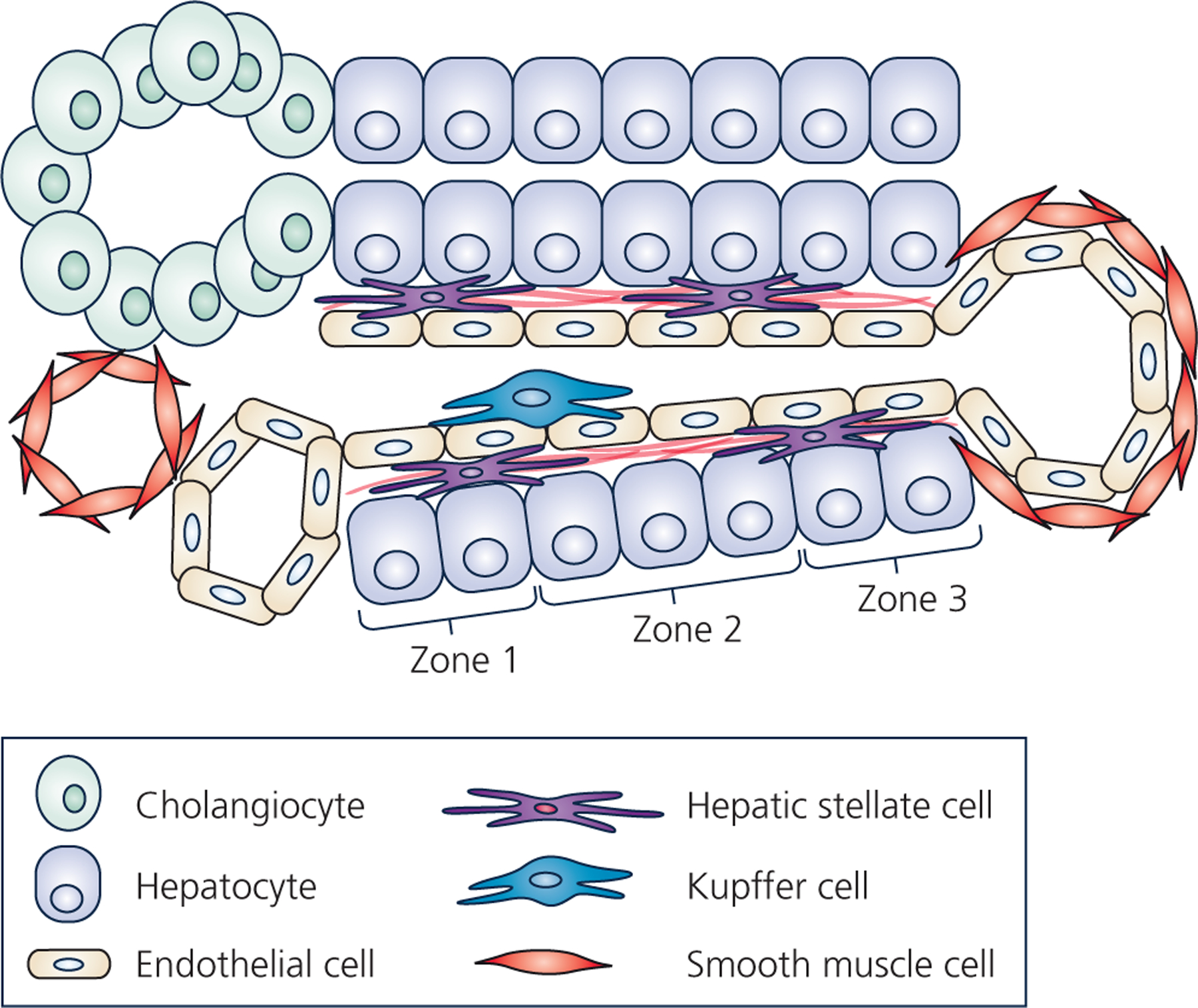
Figure 1 – Hepatic cellular architecture.

Schematic represents a cross-section of a liver lobule, which contains the complete functional structure and main cell types of the liver. One the left, is the “portal triad” comprised of the portal vein, the hepatic artery, and the bile ductule. The bile ductule is comprised of cholangiocytes (represented as green oval-shaped cells), which collects bile produced by the hepatocytes. Bile ductules ultimately combine into the bile duct which drains bile to be stored in the gallbladder. The hepatic artery (represented as a red smooth-muscle cell-lined vessel) supplies oxygenated blood originating from the celiac artery, whereas the portal vein (represented as an endothelial cell-lined vessel) supplies nutrient- and toxin-rich blood from the stomach, pancreas, gallbladder, and spleen into the liver lobule. The portal vein and hepatic artery drain blood flow into the liver sinusoid, which is lined by liver sinusoidal endothelial cells. Ultimately, blood is collected into the central vein, lined with both endothelial cells and smooth muscle cells. Resident macrophages, known as Kupffer cells (represented as blue cell), reside in the luminal side of the sinusoid. The hepatic stellate cells (HSCs) (represented as peach-colored cells with long projections) reside in the space of Disse between hepatocytes and liver sinusoidal endothelial cells and produce the basement extracellular matrix (black lines near the HSCs in space of Disse). Regions along the length of the sinusoid are commonly referred to as zones, with zone 1 being peri-portal, zone 3 being peri-central vein, and zone 2 residing between. Hepatocytes comprise the hepatic parenchyma (the predominant cell type) and display an incredible array of functions including synthesis of serum proteins, clotting factors, lipoproteins, cholesterol and bile salts, gluconeogenesis and glycogen storage, as well as detoxification. Often these different hepatocyte functions are localized to specific zones along the sinusoid.
