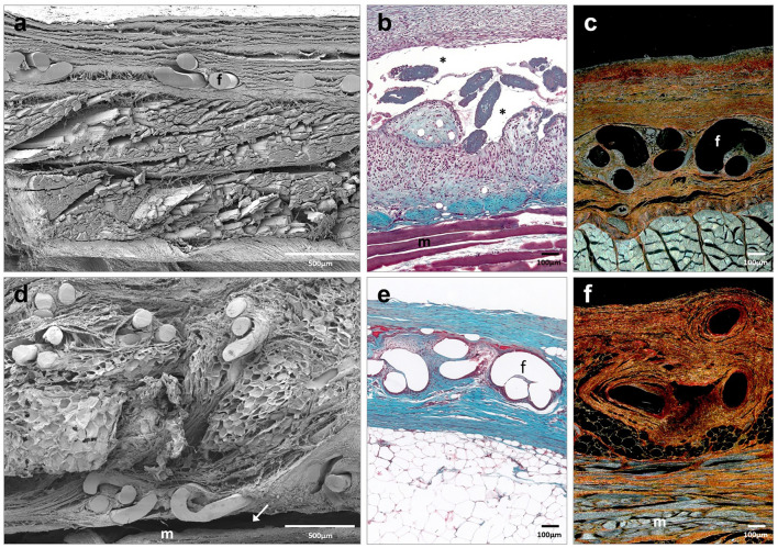
Fig. 3.
Scanning electron microscopy (a, d) (× 500; scales: 500 μm) and light microscopy images (Masson trichrome (b, e) and Sirius red staining (c, f), × 100; scale: 100 μm) of Adhesix mesh at 14 days (a–c) and 90 days (d–f) post-implantation. Sirius red staining shows collagen I (mature) in red and collagen III (immature) in yellow. Symbols: f mesh filaments; (m) muscle; (→) poor integration; and (*) area of seroma (color figure online)

