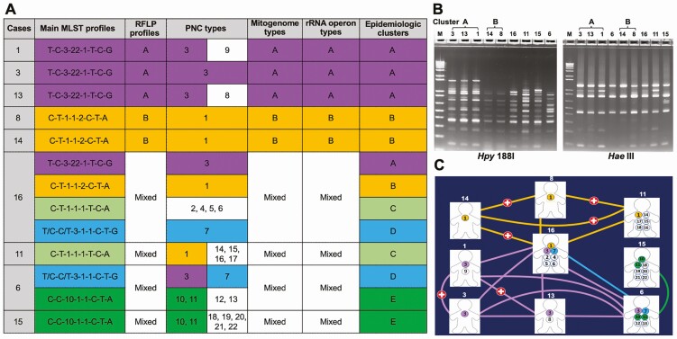
Figure 2.
Molecular epidemiology of P. jirovecii in 9 kidney transplant recipients. A, Summary of molecular genotyping and epidemiologic clusters. Each MLST profile represents a combination of SNPs (or genotypes) at mtLSU, PNC, ITS1-ITS2, 26S rRNA, soda, and dhps as detailed in Supplementary Figure 2. In cases 1, 3, 13, 8, and 14, the profiles shown represent the dominant strains. In the remaining patients (each coinfected with 4–7 strains), the profiles shown represent putative matching profiles between patients; profiles without matches are not shown to enhance visualization. Epidemiologic clusters are defined according to the consensus of the main MLST and RFLP profiles, and PNC, mitogenome and rRNA operon types. Some PNC types shared between different patients but lacking support by MLST profiles are excluded in the cluster determination. For example, PNC type 1 was shared among patients 8, 14, 16, and 11 but patient 11 did not contain a putative MLST profile shared with patients 8 and 14; thus patient 11 was not considered to be related to cluster B. Multiple strains unable to be differentiated reliably are indicated as mixed profiles or types. B, RFLP analysis of P. jirovecii samples. PCR products from the first-run were digested with restriction enzyme Hpy 188I while the semi-nested PCR products were digested with Hae III. Both gels were stained with SYBR green. Numbers at the top represent the individual patient IDs. M at the top represents DNA size markers. Samples showing the same pattern for both the first-run and semi-nested PCR products are considered the same cluster. Samples 3, 13, and 1 exhibit the same RFLP pattern (designated as cluster A) and so do samples 14 and 8 (cluster B). All the remaining samples have a unique RFLP pattern. C, Distribution of the 22 PNC types in transplant patients. Case numbers are indicated above the image of each patient; 22 PNC types (Supplementary Tables 5 and 8) are represented by numbers 1 to 22 in the circles. PNC types shared between 2 or more patients are color-coded. Patients infected with the same PNC types are connected by lines with the same colors for the PNC types. Of note, every patient shows a direct connection with at least 1 other patient, whereas patient 16 shows a direct connection with all other patients except for patient 15. Red cross indicates overlaps in clinic visit between patients. Human images are from https://www.hyglossproducts.com/people-shape-cut-outs-white-19131.html. Abbreviations: MLST, multilocus sequence typing; PCR, polymerase chain reaction; PNC, polymorphic non-coding; RFLP, restriction fragment length polymorphism; SNP, single-nucleotide polymorphism.

