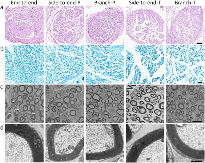
Figure 7.

Histological evaluation of the regenerated nerves 3 months after surgery (P: peroneal nerve, T: tibial nerve). (a) Hematoxylin and eosin staining (scale bar = 100 μm) and (b) Luxol fast blue staining (scale bar = 20 μm) of the regenerated nerves. (c) Transmission electron microscopy images of regenerated axons and myelin sheaths (scale bar = 10 μm). (d) Magnification of (c) (scale bar = 1 μm)
