Fig. 3.
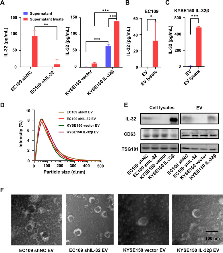
EV-IL-32 was secreted by ESCC cell lines. A Cell culture supernatant from EC109 shNC/shIL-32 (Left) and KYSE150 vector/IL-32β (Right) cell lines was treated with or without lysis buffer, and the expression of IL-32 was detected by ELISA. The EV isolated from EC109 (B) and KYSE150 IL-32β (C) were also treated with or without lysis buffer, and the concentration of IL-32 was measured by ELISA assay. D The size distribution of EV isolated from EC109 shNC/shIL-32 and KYSE150 vector/IL-32β cell lines was detected by Malvern spray analyzer. E Immunoblotting of EV markers (CD63, TSG101) and IL-32 in EV which were isolated from supernatant of EC109 shNC/shIL-32 and KYSE150 vector/IL-32β cell lines. F TEM images of EV purified from EC109 shNC/shIL-32 and KYSE150 vector/IL-32β cell lines. The images showed a mass of round-shaped vesicles. Scale bar = 100 nm. All data are representative of three independent experiments and are represented as means ± SEM. Student’s t-test, *P < 0.05, **P < 0.01, ***P < 0.001

