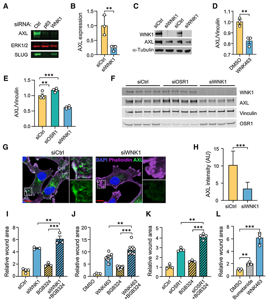
Figure 3.

Inhibition of WNK1 reduces AXL expression. A, MCF-DCIS cells (70% confluent) were treated with either control, p63, or WNK1 siRNA for 72 hours. Cells were lysed and immunoblotted for AXL, and Slug, ERK as the loading control. B, Quantification of normalized AXL expression from A; n = 3. C, SUM159 cells treated with siCtrl or siWNK1 were harvested 72 hours after transfection, and lysates were analyzed by immunoblotting with the indicated antibodies. α-Tubulin is the loading control. D, Quantification of AXL protein expression from MDA-MB-231 cells treated with WNK463 (1 μmol/L) overnight (n = 3). E, Quantification of AXL protein expression from MDA-MB-231 cells treated with control, OSR1, or WNK1 siRNA overnight (n = 4). F, Immunoblots from E. G, MDA-MB-231 cells were treated with WNK1 or control siRNAs for 72 hours. Cells were stained for AXL (green), F-actin (phalloidin, purple), and nuclei (DAPI, blue). Representative images of AXL immunofluorescence staining. Areas marked with white squares are amplified and shown in insets. Scale bars, 10 μm. H, Quantification of mean AXL immunofluorescence intensity as shown in G. An unpaired t test was performed (***, P < 0.0001; N = 60). AU, arbitrary units. I-L, Migration of MDA-MB-231 cells treated as indicated with wound area normalized to that at time 0. I, Control siRNA (n = 4), WNK1 siRNA (n = 3), BGB324 (2 μmol/L; n = 3), and WNK1 siRNA+BGB324 (n = 3) for 24 hours. J, DMSO (n = 4), WNK463 (1 μmol/L; n = 13), BGB324 (2 μmol/L; n = 10), and WNK463+BGB324 (n = 12) for 36 hours. K, Control siRNA (n = 3), OSR1 siRNA (n = 4), BGB324 (2 μmol/L; n = 3), and OSR1 siRNA+BGB324 (n = 6) for 24 hours. L, DMSO, bumetanide (20 μmol/L) and WNK463 (1 μmol/L) overnight (n = 4 for each). Data in I-L are mean ± SE; analyzed by one-way ANOVA (**P < 0.01; ***P < 0.0001). Higher concentrations of BGB324 inhibited migration more but also decreased cell viability (Supplementary Figs. S3B and S3C).
