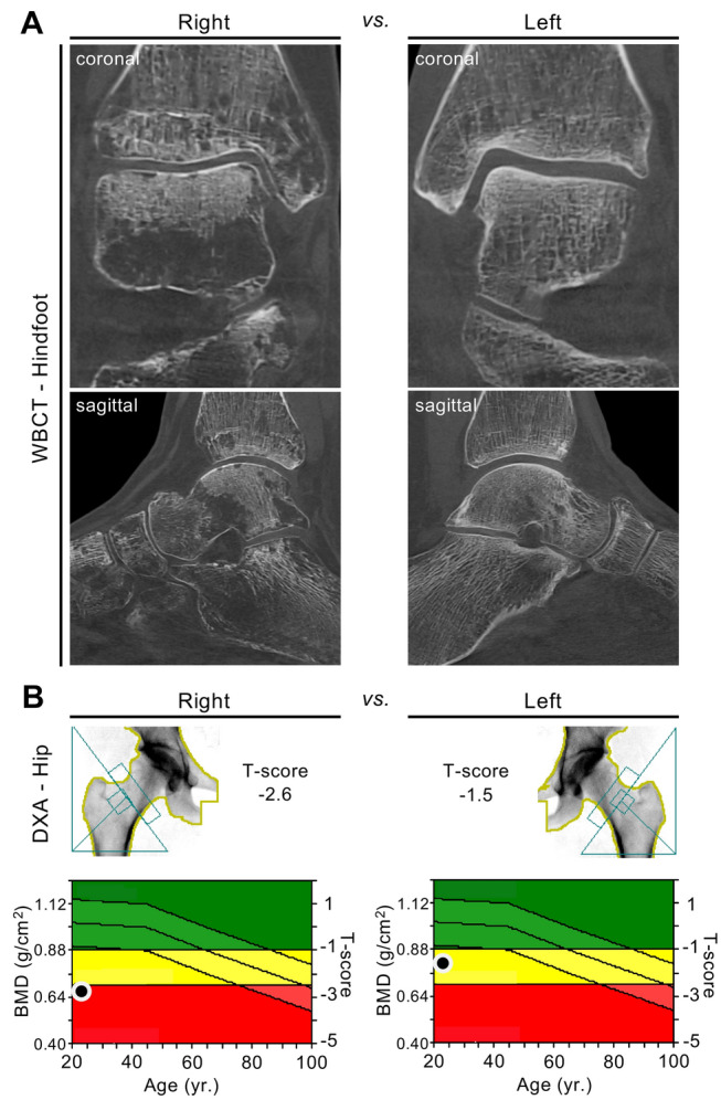
Fig. 2.

Local deterioration of bone mass after fracture and subsequent unilateral disuse of the right limb. a Cone-beam computed tomography (CBCT) images of the right foot compared to the left foot (coronal and sagittal reconstruction) in a 22-year-old female patient obtained eight months after suffering from an ankle fracture with subsequent unilateral unloading of the right limb. Focal osteolytic changes (often referred to as “local disuse osteopenia/osteoporosis”) are visible. b DXA scans demonstrating the differences in BMD between the right and left proximal femur (T-score − 2.6 vs. − 1.5). Bilateral HR-pQCT scans of the distal tibiae were also performed and showed cortical and trabecular bone loss syndrome on the unloaded side compared to the contralateral (loaded) side (cortical thickness − 28%, bone volume per tissue volume − 7%)
