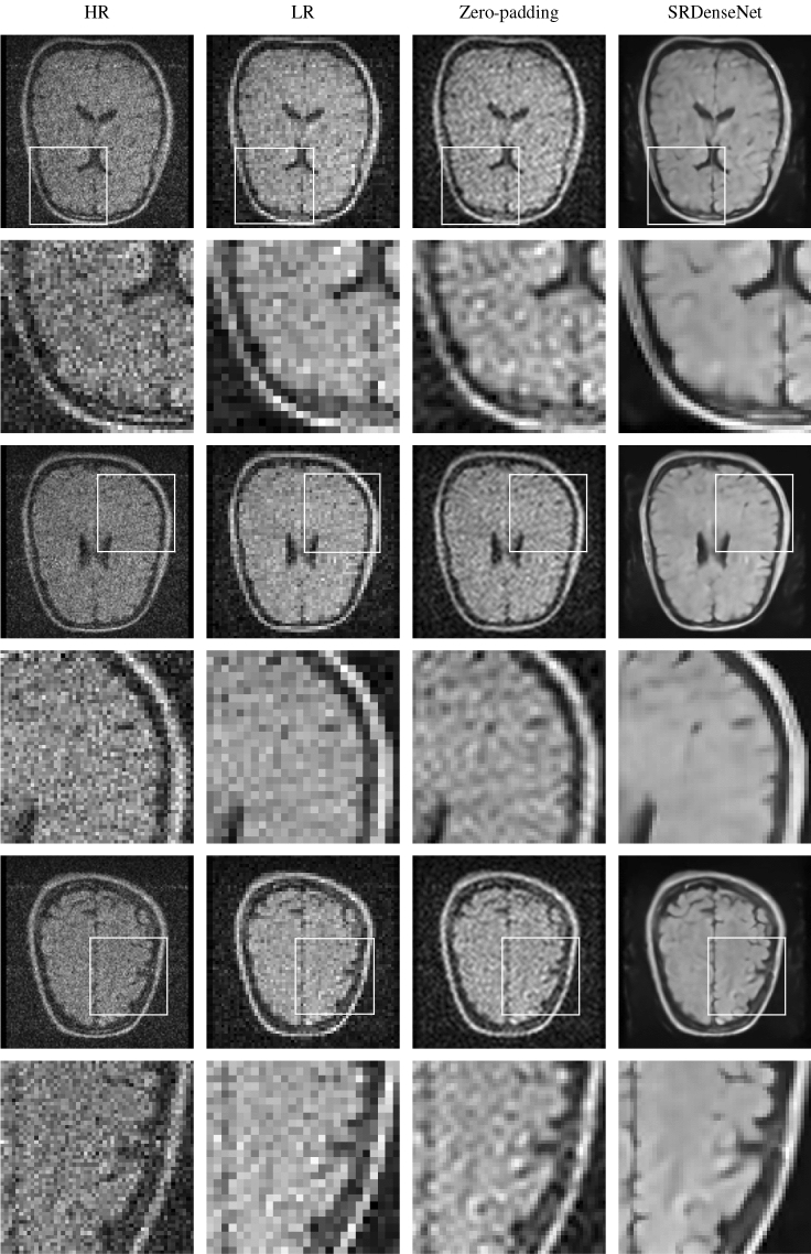
Figure 6.
In the first column, we have three different HR slices of a second brain image acquired using a low-field MRI scanner. Again, these HR images are our reference images. The first, third and fifth rows show the full images, in the second, fourth and sixth rows we find zoomed-in versions of patches of the images in the first, third and fifth rows. The second column shows the LR images corresponding to the HR images in the first column. These images are fed into the trained convolutional neural network. The third column shows the SR images obtained by zero-padding k-space, and in the fourth column, we see SR images obtained by applying SRDenseNet.

