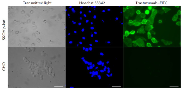
Fig. 3.
Imaging of the HER2 receptor expression in SKOVip-kat (HER2-positive) and CHO (HER2-negative) cells using the monoclonal antibody trastuzumab conjugated to the fluorescent dye FITC. Expression of HER2 on the SKOVip-kat cell surface was confirmed by intense staining of the cell membrane with the anti-HER2 antibody. Cell nuclei were stained with Hoechst 33342. The excitation and emission wavelengths for fluorescence detection were as follows: 405/10 and 460/40 nm Hoechst 33342 and 470/40 and 525/50 nm for FITC, respectively. Scale: 50 μm

