Fig. 7.
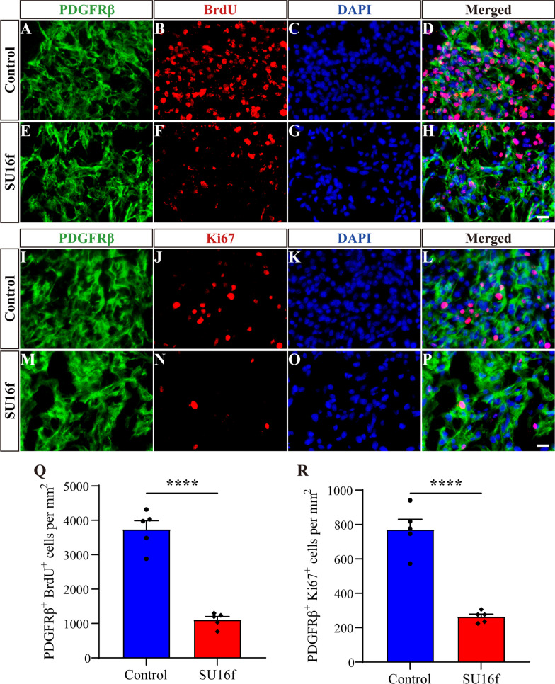
Intrathecal injection of SU16f inhibits fibroblasts proliferation after SCI. A–H Immunofluorescence staining of BrdU (red), PDGFRβ (green) and nuclei (blue) in sagittal sections of the Control and SU16f groups at 7 dpi. I–P Immunofluorescence staining of Ki67 (red), PDGFRβ (green) and nuclei (blue) in sagittal sections of the Control and SU16f groups at 7 dpi. Q–R Quantification of the density of BrdU+PDGFRβ+ cells Q or Ki67+PDGFRβ+ cells R at 7 dpi. Scale bars: 20 μm. ****P < 0.0001 by Student’s t test, n = 5 animals per group

