Fig. 8.
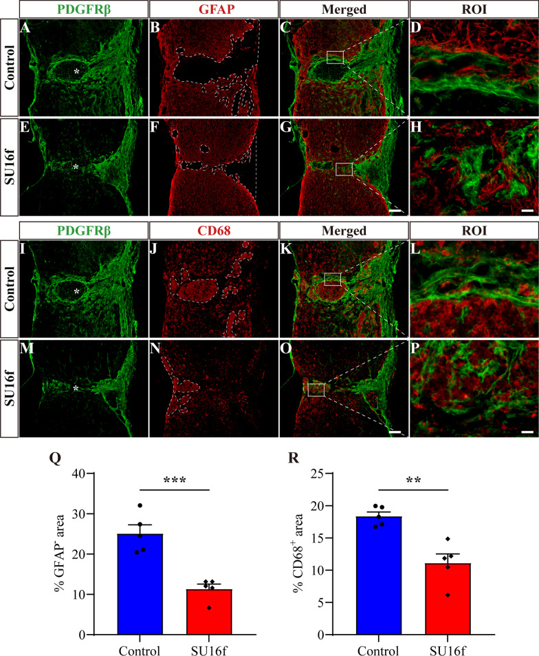
Intrathecal injection of SU16f breaks the scar boundary, inhibits the lesion and inflammation after SCI. A–H Immunofluorescence staining of GFAP (red) and PDGFRβ (green) in sagittal sections of the Control and SU16f groups at 28 dpi. The region of interest (ROI) represents the boxed region on the left and shows the fibrotic/astrocytic scar boundary. I–P Immunofluorescence staining of CD68 (red) and PDGFRβ (green) in sagittal sections of the Control and SU16f groups at 28 dpi. ROI represents boxed region in the left. Q–R Quantification of the percentage of GFAP− area Q or CD68+ area R in the area of the spinal cord segment spanning the injured core at 28 dpi. Asterisks indicate the injured core. Scale bars: 200 μm in G and O and 20 μm in H and P. **P < 0.01 and ***P < 0.001 by Student’s t test, n = 5 animals per group

